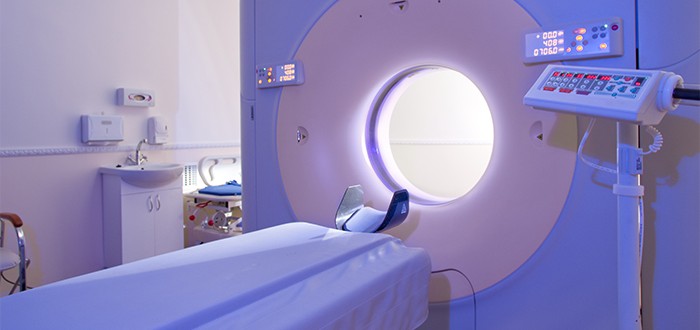
Using radiation tomotherapy following pleurectomy/decortication surgery can help keep mesothelioma away from the treated area longer than can standard three-dimensional conformal radiation therapy.
Researchers from the David Geffen School of Medicine at the University of California, Los Angeles reported this finding in the journal Practical Radiation Oncology.
The researchers observed that tomotherapy improves local control over advanced malignant pleural mesothelioma. They also observed that tomotherapy changes localized failure patterns in ways favorable to the patient.
With tomotherapy, the median time until local control over the mesothelioma is lost after pleurectomy/decortication surgery measures 19 months, the researchers wrote.
With 3D conformal radiation therapy, the median time until loss of local control is a much shorter 10.9 months, they indicated.
The two methods of delivering radiation were evaluated with the help of 45 study patients, all with advanced malignant pleural mesothelioma.
Tomotherapy Works Well After Pleurectomy/Decortication Surgery
The researchers said they made these observations while testing their hunch that helical tomotherapy might be superior to 3D conformal radiation therapy when administering intensity modulated radiation.
They were particularly keen about testing to see whether that superiority extended to improving local control of advanced malignant pleural mesothelioma after a pleurectomy/decortication surgery.
Intensity modulated radiation is customarily administered following the surgery in order to knock down mesothelioma cells that the knife missed and left behind.
The shortcoming of the conventional method of delivering intensity modulated radiation is that it kills with too broad a sweep. In other words, it has the ability to knock down too many healthy cells and tissues right along with the leftover mesothelioma cells.
Consequently, radiation oncologists cannot administer the therapy too aggressively. Unfortunately, going easy on the radiation dosing allows the mesothelioma to recur relatively soon.
In contrast, tomotherapy allows doctors to administer stronger doses of radiation. The reason they can do that is because the technology allows for delivery with far greater precision.
Since the radiation is more precisely delivered, there is less risk of knocking down healthy neighboring cells and tissues. Avoiding harm to healthy neighboring cells and tissues is important because their survival means fewer manifestations of the unpleasant side effects of radiation.
And hitting the remaining mesothelioma cells with the highest possible dose of radiation is important because it helps ensure that they won’t recur so soon.
Tomotherapy Targets Mesothelioma Cells
The proton radiation delivered by a tomotherapy machine is produced using a linear accelerator. As the radiation is emitted, it passes as a single beam through a unique multileaf collimator.
The multileaf collimator splits the beam into multiplied thousands of needle-thin beams. Special software calibrates the intensity of each individual beam and directs it to a precise target.
The target is identified at the start of the treatment session. Identification is accomplished by taking a CT image of the chest region with the aid of the machine’s built-in scanner.
The image shows exactly where the mesothelioma cells are located. The image is also in 3D, so the full width, length and height contours of the mesothelioma cells are revealed.
The software uses this image as a basis for calculating the individual beam strengths that are required to get the job done. The software also takes into account physician-supplied treatment parameters.
The tomotherapy also permits the radiation to be administered from any direction. This is possible thanks to the machine’s ring-shaped gantry.
All of these features work together to help ensure that any remaining mesothelioma cells stay knocked down for the longest time possible following mesothelioma pleurectomy/decortication surgery.

